91 Animal Ultrasound Images
This site uses cookies By continuing to browse this site you are agreeing to our use of cookies. We have created an online library of small animal veterinary images and movie clips to help you learn more.
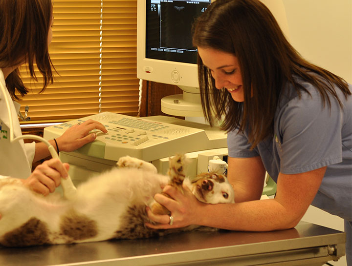
Veterinary Ultrasound Dog Ultrasound In Salem
Ultrasound imaging sometimes referred to as sonographic imaging involves the use of sound waves to image the organs in the patient.

Animal ultrasound images. Fluorescent Imaging Using CRi Maestro. Many studies are performed to examine tendons ligaments and joints as part of the comprehensive evaluation of horses with lameness or performance problems. Oblique ultrasound image near the right cranial quadrant in a cat B.
Ultrasound-guided biopsy and endoscopic procedures 60 to 90 minutes A copy of the report is faxed to your veterinarian and is also made available to you either at the conclusion of the office visit or by mail or email within 24 hours. Several types of image formats can be displayed. Ultrasound is a non-invasive immediate tool used to image tissue.
The Large Animal Ultrasound Service offers a complete range of ultrasound imaging procedures. Diagnostic ultrasound uses the high frequency sound waves to make images of the bodys internal organs much like a high detail version of the sonar images used by fisherman for detecting fish. 23 rows Radiology is a medical specialty that focuses on using advanced imaging technologies such.
Reading animal ultrasound images EI. The bile duct 2 mm can be visualized in this cat arrow. We partner with your veterinarian to provide you with the information you need to make informed choices about your pets healthcare.
So the first step to help you read the ultrasound image is. All of the images are stored on a central computer allowing the images to be viewed on all computers in both hospitals. - vet ultrasound stock pictures royalty-free photos images.
B B. Animal Ultrasound Services is an outpatient veterinary imaging center that provides diagnostic ultrasounds fine needle aspirates and clinical consultations for dogs cats and exotic species. The caudal lung field part of the liver the elongated kidney and the cigar-shaped active right testis.
It allows visualizing a longer region of interest. Small animal image library. Three transducer port is designed to meet the different clinical applications.
It uses ultrasonic sound waves in the frequency range of 1515 megahertz MHz to create images of body structures based on the pattern of echoes reflected from the tissues and organs being imaged. Versatile and easy to use. The cystic and bile ducts will not be dilated in the normal dog.
Ultrasounds are safe and radiation-free so they are harmless for people and animals alike. Small Animal Ultrasound the capability to extend ultrasound imaging to mouse rat and other animal models with excellent spatial and temporal resolution. Here are some optical and ultrasound images from the Small Animal Imaging Resource at UT Southwestern UT-SAIR.
An ultrasound is a form of diagnostic imaging that uses sound waves to create an image called a sonogram of the inside of the body. Davidianus in panoramic view mode consisting of several single ultrasound images. Small animal ultrasound and X-ray Image library.
Veterinarian with assistant examining dog in animal hospital - vet ultrasound stock pictures royalty-free photos images above view of team of veterinarians having an ultrasound exam on a doberman at animal hospital. It will not penetrate bone like an X-Ray. Latest design proven solutionall veterinary application 15 inches color LED touch screen with 45 degree tilt functionality.
Novel pH sensitive nanoprobe for detecting tumors growing in mice breast and brain cancers shown. From left to right. If you are considering entering this field make sure to do more research on colleges or universities that will allow you to get hands on work with the actual equipment.
Preclinical Imaging the benefits of modern dignostic medical imaging systems to the studies of anatomy and physiology in small animals. Ultrasonography is the second most commonly used imaging format in veterinary practice. This is a normal finding.
Long-axis right-sided image of the liver and gallbladder in a normal dog A. Ultra pH-sensitive fluorescent nanoprobes for cancer detection. Medical Imaging a world leader and manufacturer of rugged portable ultrasound for the veterinary and livestock industry.
Small Animal Digital Radiology The Radiology Service in the Wilford and Kate Bailey Small Animal Teaching Hospital provides digital radiography for teaching research and patient evaluation. G Sonogram of an adult male giant salamander A. Advanced imaging technology and superior image quality can provide fast and precise scans.
By obtaining images of their internal organs the ultrasound techs can help the doctor or vet make a good diagnosis ensuring that the animal will get the proper treatment.
